
NEWS
Nikon Reopens a Beauty Contest Winner Over AI Color
Nikon is re-reviewing Ning Xu’s winning cilia video after biologists said AI coloring invented cell structures the contest had sold as diagnosis.

Nikon is re-reviewing its 2026 Small World in Motion winner after biologists said an unsupervised network painted structures that living airway cells do not have. Dr. Ning Xu’s clip of beating cilia still sits in first place. The caption now calls it a rendition.
The fight is not a simple cheat-or-not story. Nikon scored the contest on artistic impact, sold the movie as disease diagnosis, and wrote a rule that only bans generation.
Nikon Called the Clip a Diagnosis, Then a Rendition
On September 15, 2026, Nikon Instruments named Xu the winner of the 16th annual video contest. He was listed then as an optical engineer in the Department of Precision Instrument at Tsinghua University in Beijing. Nikon later said he had joined the National University of Singapore as a research fellow.
The press note did not whisper. It said the movie showed why advanced microscopy matters in the diagnosis of diseases defined by movement. Primary ciliary dyskinesia, or PCD, is a rare genetic disorder in which airway cilia beat the wrong way, so mucus and debris stay in the lungs. Xu’s line was blunt: to understand the disease, you need to watch about 10 seconds of motion, not a still frame.
Eric Flem, senior manager of communications and CRM at Nikon Instruments, said the clip showed why the company added a video category 16 years earlier. Motion, he said, can change how people understand the microscopic world. The official account posted the winner with a gold medal and a link to a profile essay.
🥇 Congratulations to Dr. Ning Xu for winning this year's Nikon Small World in Motion competition!
Read more about Dr. Ning Xu and his winning video in his Masters of Microscopy article: https://t.co/5fAIQZg8bY pic.twitter.com/JuU4sZs2fa
— Nikon Small World (@NikonSmallWorld) September 17, 2026
Seven days later the profile essay changed. Nikon wrote that an unsupervised neural network had been used in post-processing and visualization, after grayscale reconstruction, “to distinguish and visualize features in the grayscale data.” By September 24 the gallery title itself had shifted. The page now heads the clip as a rendition of abnormal beating of airway cilia, with a line that it was AI-assisted in post-processing.
HOW NIKON NAMED THE SAME FILE
| Date | Public language |
|---|---|
| September 15, 2026 | First place, abnormal beating, framed as diagnosis through motion |
| September 22, 2026 | Masters essay adds an unsupervised network used after grayscale reconstruction |
| September 24, 2026 | Gallery title becomes a rendition, with an AI-assisted post-processing note |
| September 26, 2026 | LinkedIn: carefully re-reviewing vetting files and new technical documents |
A Nikon spokesman had already told researchers the enhancement tool was being treated as processing of microscope data, not generation from scratch, and as inside the rules. After the pushback, the same office said it was reviewing the AI policy. “At this time, we don’t see that any rules were violated, but we are reinvestigating and re-evaluating the entry.”
The Child’s Cilia Had to Be Filmed Alive
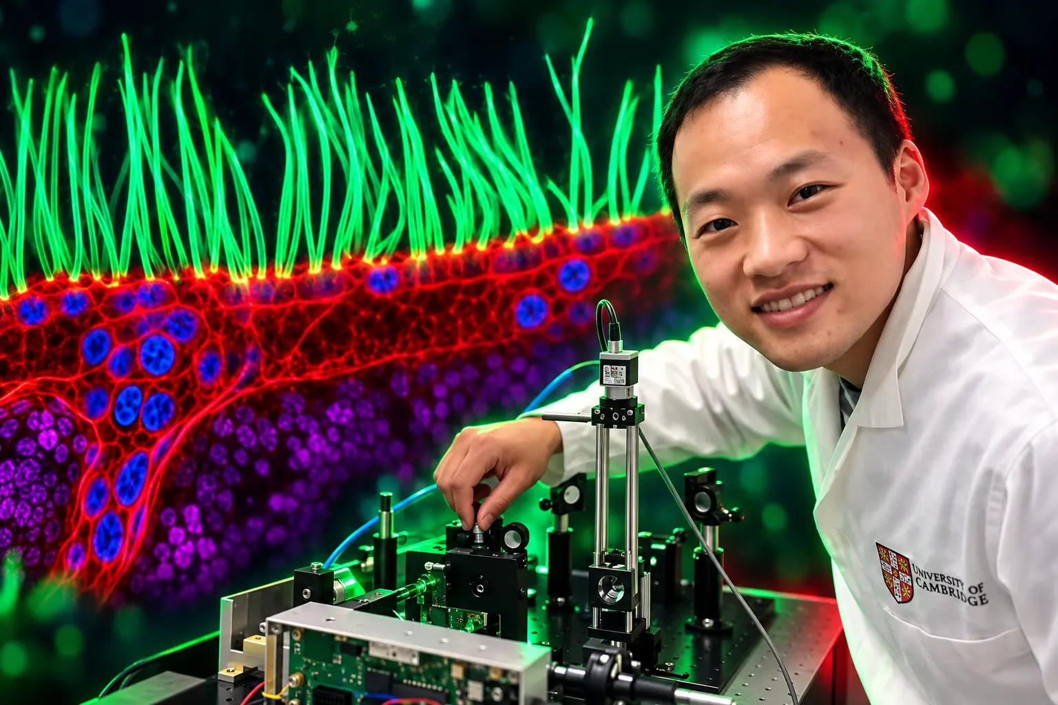
The sample was patient-derived airway tissue from a child with PCD. Xu’s problem was physical. Cilia are small, they beat fast, and the tissue has to stay alive and behave as it would in the body. Many high-end methods lean on fluorescent labels or hard light, which can change or damage living cells.
He and his collaborators shaped several light waves with a digital micromirror device, keeping the dose down while still grabbing a high-resolution moving image. Nikon lists the method as diffractive super-resolution microscopy through a 100X objective. The Masters essay says the winning run was recorded in December 2025 on the fifth version of a system whose development started in 2021.
THE CAPTURE, IN NIKON’S OWN FIGURES
- Lens: 100X objective, diffractive super-resolution on a custom optical bench.
- Recorded: December 2025, on the fifth version of a build begun in 2021.
- Watch window: Xu said about 10 seconds of motion is what the disease requires.
- Color step: an unsupervised network run on reconstructed grayscale frames, after the movie existed.
That last step is the whole argument. Xu has said, more than once, that AI did not generate the experimental movie, the cilia, or their motion. Color and separation, he said, came later, on data the microscope had already produced.
Biologists Count About 200 Cilia, Not One
Robert Hirst, a lead scientist who uses microscopy to diagnose PCD at the University of Leicester, did not wait for a watermark debate. “When you start looking at the scales of the movie and some of the details of it, it was quite clear to the community…that some of these features just are not consistent with nature,” he said.
Edward Phelps, an associate professor at the University of Florida, wrote that there were “many serious problems with this video,” and that “the microscopy community deserves an explanation from Nikon.” His list was anatomical, not aesthetic.
WHAT CRITICS SAY THE CLIP GETS WRONG
- The purple bodies: Phelps said they look like mitochondria, but a sub-epithelial pile of extracellular mitochondria the size of nuclei does not occur in biology.
- The blue bodies: they resemble nuclei in stills, he said, and then fail to behave as nuclei in motion.
- The red channel: Phelps said it is unclear what the red stain is supposed to be.
- The count: Hirst said the film reads as one cilium per cell; a real airway cell carries about 200.
- The floor: structures “pop in and out of existence,” cilia “appear from nowhere,” and the layer under the hairs is, in Hirst’s words, “wrong in scale, wrong in biology, and just looks completely fabricated.”
What worries me most of all is that PCD patients will see this video and think, ‘My cilia look like this, and my cells look like this,’ and they just don’t, unfortunately.
Robert Hirst, lead scientist, University of Leicester
Christophe Leterrier, a neuroscientist at CNRS and Aix-Marseille University and a former Small World juror, put the same charge in contest language. The processing, he said, altered the original image too much. “You have something very nice and very engaging visually but then it’s absolutely not representing a biological reality.”
Melanie White, a developmental biologist at the University of Queensland, refused the idea that a contest still is only a picture. “Scientific images are not just illustrations,” she said. “They are data, and we need to be able to trust that what we are seeing is grounded in the underlying measurement.”
Xu answered on Nikon’s LinkedIn post. The video was made for a contest that celebrates the beauty of microscopy, he wrote, so the team wanted it visually engaging as well as scientifically interesting. Post-processing, he said, enhanced the regions below the cilia “without making anatomical claims about what those rendered features represent.”
For me, the important distinction is between using an image to make a scientific claim and using visualization to communicate the beauty of scientific data. Both can have a place, as long as we are clear about which is which.
Ning Xu, comments on Nikon’s LinkedIn post
That distinction would be cleaner if Nikon’s own winner note had not sold diagnosis. It would also be cleaner if the gallery had opened as a rendition instead of arriving there after specialists started counting cilia.
The Rulebook Stops at the Word Generated
The posted rules for the next cycle, with entries open from July 24, 2026, through April 30, 2027, say AI generated videos are not permitted and that Nikon may demand the original file. Another line voids submissions “generated by AI, script, macro or other automated means.” Nowhere does the text define enhancement, false color, reconstruction, or an unsupervised network.
Dr. Patrick Hickey, who took fifth with a confocal movie of mitochondria and chloroplasts in Turtle Vine leaf cells, said the 2026 contest already barred generative AI and required a light microscope. “There’s a specific set of rules in the contest, and one of them was quite clearly that you couldn’t use generative AI to produce things, and the other was obviously that it has to be taken under a microscope,” he said.
Judges are told to score creativity and originality, informational content, technical proficiency, and artistic and visual impact. Xu wrote the sentence the artistic line invites. “Embrace AI to reveal the hidden beauty in your data,” he advised other researchers in the Masters essay.
Markus Sauer, who studies super-resolution microscopy at the University of Würzburg, said using AI to visualize experimental data is not automatically a problem. It becomes one, he said, when the result misrepresents, exaggerates, or alters the finding. White drew the same split in plainer words: enhancing information that was already captured is one act, and creating information that was not there is another. “It’s difficult to understand where Nikon is drawing the line around the use of AI,” she said.
Leterrier said the field’s objection is not to AI as such. Microscopists already lean on it. The objection is the extent of the change, and the fact that the extent was not on the label when the gold medal went up.
What a SynthID Watermark Would Mean
Ian Donovan, a PhD student at UT Southwestern Medical Center, wrote that the file carries “an embedded SynthID watermark identifying synthetic/AI-generated content.” He described running the clip through Google Gemini and getting that reading back.
Google’s own pages present SynthID as an invisible watermark in AI-generated video written into pixels when its models create a frame. The consumer stack named on those pages is Gemini, Imagen, Lyria, and Veo. A detector can highlight which regions of a file are most likely marked. Absence of a mark never rules generation out, because other tools do not write SynthID.
That limit cuts both ways. A custom unsupervised network trained in a university lab would not ordinarily stamp Google’s mark. If Donovan’s reading is right, some Google generative tool touched at least part of the pixels, which is a narrower claim than “AI was used in coloring.” If the reading is wrong, the watermark is a sideshow and the anatomy fight remains.
Xu has promised a paper with a fuller description of the optical system and a quantitative analysis of the ciliary motion. Until that package is public, Nikon is holding technical documents that the rest of the field has not seen.
A Fifth-Place Filmmaker Wants the Same Rules for Everyone
First prize is $3,000. Second is $2,000, third $1,000, fourth $800, fifth $600, with honorable mentions at $200. Hickey said the recognition matters more than the check, and that it would be unfair on other entrants if the winner had been altered in a way the rules did not allow.
Dr. Andrew Moore of the Howard Hughes Medical Institute’s Janelia campus, who placed fourth with synchronized cell division, called Xu’s admission that regions under the cilia were rendered “startling.” Valentin Dunsing-Eichenauer, a researcher at Der Simulierte Mensch in Berlin, called the clip a misuse of AI in bioimage post-processing.
The early audience for the winner post was not that jury. Replies treated the clip as a national science win: a Tsinghua-trained engineer, a custom microscope, a rare disease made visible. The second audience watched nuclei that do not act like nuclei. Some cell biologists now want the medal pulled, and at least one has urged a professional society to walk away from Small World if it stays in place. Nikon has not taken the file down.
Xu is, in the company’s phrase, “respectfully complying.” He has sent equipment lists, imaging methods, and processing notes. Nikon said its team at first found those methods “technically legitimate and consistent with established microscopy practices.” Established practice is the soft ground. False color, deconvolution, and reconstruction were already how living cilia get shown. An unsupervised network made the old habit watermarkable, and made the pretty floor under the hairs look like a claim.
Journals Already Require Authors to Name the Processing
A contest is not a paper, which is Xu’s best defense. It is also why the gap looks sloppy. Journal figure guides already say generative AI in figures is not permitted, including content-aware Photoshop tools. They tell authors to disclose pseudo-coloring, gamma changes, deconvolution, and rendering, and to keep the final image a fair record of the original data.
Clinicians who actually diagnose PCD do not need a beauty pass. The National Heart, Lung, and Blood Institute lists video microscopy of airway cilia as one of the tests: a brush sample, a microscope, and a look at how the hairs move. Hirst’s fear is that a contest clip labeled as a child’s disease will be read as that test.
The 2026 judging panel was Joe Dragavon of the University of Colorado Boulder, Quinten Geldhof of the Museum of Science in Boston, science writer Corey S. Powell, and Meredith Sagolla of Genentech. Videos may run no longer than 60 seconds, must come from a light microscope, and may not carry sound or graphics. Small World itself is 50 years old in Xu’s telling. The motion contest began in 2011.
Xu still has the first-place line on the gallery. Nikon still has a rule that names generation and does not name color. The clip that was supposed to show a child’s broken cilia is, for now, a rendition of that idea, under review, with the medal where the company left it.

Greed Held After a $697 Million Crypto Long Flush

Fed Minutes Keep a Year-End Rate Hike Alive

A Nanny-Cam Tape Follows Tyreek Hill Into Free Agency

Star Wars Galactic Racer Makes a Crash End the Tour

Amazon Puts Apple’s June MacBook Prices Back on Sale

Sophia Wootten Turns the Miss Georgia Crown Toward Cleft Care

Georgia Beach Bacteria List Leaves Swim Areas Open

The iPhone 16 Pro Flipkart Cut Did Not Last

Little St. Simons Island Hatches Loggerheads at 72.4 Percent

IXI Autofocus Glasses Still Lack a European CE Mark
-
 ENTERTAINMENT4 months ago
Sophia Wootten Turns the Miss Georgia Crown Toward Cleft Care
-
 NEWS4 months ago
Georgia Beach Bacteria List Leaves Swim Areas Open
-
 NEWS1 year ago
The iPhone 16 Pro Flipkart Cut Did Not Last
-
 NEWS3 months ago
Little St. Simons Island Hatches Loggerheads at 72.4 Percent
-
 NEWS10 months ago
IXI Autofocus Glasses Still Lack a European CE Mark
-

 BUSINESS2 months ago
UltraTech’s Ultravolt Launch Puts Cable Multiples on Trial
-

 NEWS2 months ago
Microsoft Prints Azure’s $29.42 Billion Quarter and Shrinks the Map
-

 NEWS3 years ago
Eyal Waldman Keeps a Two-State Bet After Gaza